
Dosimeter badge services for medical, dental, and veterinary businesses
Learn how Radiation Detection Company’s easy-to-use dosimetry solutions can boost the efficiency of your practice.
Since its discovery in 1895, radiation has revolutionized both science and medicine, although early researchers had no idea the dangers it presented. In this article, we'll dive deeper into the history of radiation (including its discovery and major inventions), in addition to exploring the evolution of radiation safety practices such as x-ray badges.
On November 8, 1895, a German physicist named Wilhelm Conrad Röntgen was experimenting with electric current flow in a partially evacuated glass tube (cathode-ray tube). Röntgen observed that a screen of barium platinocyanide far away from the tube gave off light when the tube was operating. He theorized that when the cathode rays (electrons) struck the glass wall of the tube, some unknown radiation was formed. The unknown radiation then traveled across the room, struck the chemical, and caused the fluorescence. Later investigations revealed that other materials (including paper, wood, and aluminum) were transparent to this form of radiation. He mistakenly thought the rays were unrelated to light, and due to its uncertain nature, he called the phenomenon x-radiation.
Röntgen took the first x-ray photographs of the interiors of metal objects and the bones in his wife’s hand. He did not patent his discovery (which also became known as Röntgen radiation), because he wanted his work to benefit all. In 1901, he received the first Nobel Prize for Physics for his work on x-ray radiation, and donated the money he received to the University of Würzburg (where he had accepted an honorary doctorate of medicine). The use of x-rays in medicine—for both medical imaging and the treatment of cancers—was almost immediate, happening only weeks after the announcement of Röntgen’s discovery in early 1896.
Marie Curie shared the Nobel Prize for Physics in 1903 with Henri Becquerel for their contributions to the understanding of radioactivity. Working with her husband Pierre, they discovered polonium and radium. After her husband's death, Marie's devotion to developing the use of x-radiography only strengthened. She played an integral part in the completion of the laboratories at the Radium Institute at the University of Paris, which would go on to become a universal center for chemistry and nuclear physics.
Marie Curie's death by aplastic anemia in 1934 was caused by the action of the radiation she studied for over thirty years. Nevertheless, her influence in nuclear physics and chemistry was immense and impacted many generations to come.
As we mentioned above with Marie Curie, many early pioneers in radiation research suffered severe health issues, with many cases resulting in death. Notable victims included Thomas Edison's assistant, Clarence Dally, who ultimately lost both of his arms due to high levels of radiation exposure, and the "Radium Girls."
The "Radium Girls" were a group of female factory workers who painted watch dials with radioactive paint. Their work led to severe radiation poisoning in many workers, resulting in more than thirty deaths due to radiation exposure. These high-profile examples (media coverage and litigation for "The Radium Girls" was plentiful) led to the emergence of radiation protection regulations and guidelines. The formation of the International Commission on Radiological Protection (ICRP) in 1928—and later in the 1970s, national regulatory bodies like the US Nuclear Regulatory Commission (NRC) and the UK's Health and Safety Executive (HSE)—proved to be vital steps to combat radiation exposure.
In 1928, Hans Geiger (a renowned German physicist) and Walther Müller (a PhD student of Geiger) developed the sealed Geiger–Müller tube. This small device used basic ionization principles to detect alpha and beta radiation as well as gamma radiation (which prior models had been unable to do). This practical instrument could be produced relatively cheaply, as the tube output required little electronic processing. Due to minimal valve count and low power consumption, the Geiger counter (as it became known) achieved great popularity as a portable radiation detector. To this day, the Geiger counter is a staple in many industries used to detect ionizing radiation.
A Scintillation Detector (or Scintillation Counter) is an instrument used to detect and measure ionizing radiation that uses the excitation effect of incident radiation on a scintillating material and detecting the resulting light pulses. The first electronic scintillation counter was invented in 1944 by Sir Samuel Curran at the University of California at Berkeley. A requirement to measure the radiation from small quantities of uranium birthed this innovation, which used the newly available and highly-sensitive photomultiplier tubes to accurately count the flashes of light from a scintillator subjected to radiation.
Shielding is one of the three main tenets of radiation safety (more on this below). Shielding relies heavily on the use of Personal Protective Equipment (PPE) for radiation protection, which includes lead aprons or vests, lead thyroid collars, lead gloves, and safety glasses (as the lens of the eye is one of the body's most radiosensitive tissues). PPE is commonly used in settings where medical radiation is necessary. For more on this, please check out our guide on materials that block radiation.

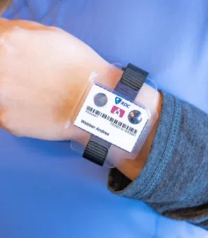
A radiation badge—also commonly called a personal dosimeter—is a device that measures the absorbed dose of ionizing radiation for personnel that are in occupational contact with radioactive material. Radiation monitoring badges are the main component of radiation dosimetry, which is the accurate and systematic measurement, calculation, and assessment of the ionizing radiation dose absorbed by matter or tissue.
Dosimetry is broken into two branches: internal absorption and external radiation. X-ray badges are crucial to external radiation. The introduction of dosimeters was vital in the tracking of occupational radiation exposure, and subsequently improved workplace safety for radiation workers in various fields (including nuclear power plants). Normally those in contact with radioactive substances during the regular course of their employment (or those who have the potential to be exposed to radiation), carry personal x-ray badges somewhere on the outside of their clothing. Continued technological advancements and constant regulatory compliance are paramount to the safety of radiation personnel.
The earliest form of personal dosimeters, film badges used photographic emulsion to measure radiation exposure. Early film x-ray badges were not very sensitive, and easily impacted by adverse environmental conditions like temperature and humidity. They also had to be exchanged every two weeks to be processed and analyzed. Results were recorded on index-sized cards called dosimetry cards. These cards were used to record an employee’s cumulative external exposure to radiation, and this process continued until the late 1970s. In 1979, an early computer system replaced dosimetry cards. Film badges were replaced in 1994 by Thermoluminescent Dosimeters (TLD).
TLD dosimeters provided a more accurate and reliable measurement of occupational dose than their film badge predecessor. As ionizing radiation passes through a thermoluminescent dosimeter, electrons in the material are moved into dosimetric traps. The electrons are held there until the detector is heated up. Once the temperature reaches a maximum of approximately 400° Celsius, the trapped electrons begin to move. This movement causes a light pulse to be emitted, also known as luminescence.
In luminescence, only a small fraction of atoms (called the emission center or luminescence center), emit light. A photomultiplier tube (PMT) is then used to count the visible light emitted. The amount of emitted light is proportional to the amount of ionizing radiation exposure that the dosimeter received.

In 1998, Optically Stimulated Luminescence (OSL) Dosimeters were introduced. The material makeup of a TLD dosimeter and an OSL dosimeter is very similar, with both composed of crystalline solids. Both dosimeters capture point dose measurements, or radiation doses in a relatively small volume.
An OSL dosimeter most commonly uses Beryllium Oxide (BeO) to absorb x-ray energy. It then releases it and measures the precise dose of ionizing radiation received. Beryllium Oxide is commonly used because it is extremely durable, sensitive, and resistant to environmental influences and fading. Some OSL dosimeters use Aluminum Oxide (Al2O3:C) instead of BeO.
An electronic personal dosimeter provides a real-time direct display of information about the measured dose for the individual wearing the device. This real-time reading enhances radiation worker safety (ensuring levels don't exceed body dose limit) and enables for a quicker response to potential hazards.
The goal of any radiation safety program is to keep radiation doses as low as reasonably possible ("ALARA"). There are three basic protective measures to minimize radiation exposure are:
Time refers to the duration spent near a radiation source. The amount of time should be minimized whenever possible, while of course still allowing the worker to accomplish necessary tasks.
Distance refers to the proximity to a radiation source. The amount of distance should be maximized whenever possible. Increasing distance leads to decreasing dose.
Shielding refers to simply putting something between you and the radiation source (whether it be a wall, protective clothing, or other equipment to assist in proper shielding).
Radiation Detection Company has 75 years of experience providing quality dosimetry service to over 28,000 companies nationwide. Our long history of safety and innovation makes us the cornerstone of an affordable radiation safety program for organizations of all sizes and industries. Need help understanding what dosimeters your organization needs? Please contact us, and our team will be happy to provide guidance.
Need a question answered that we did not address in this article? Please reach out to our Customer Care team, and one of our specialists will be more than happy to help.
Learn how Radiation Detection Company’s easy-to-use dosimetry solutions can boost the efficiency of your practice.
Radiation safety standards vary widely from state to state. To help you determine the requirements for your practice, we’ve put together this comprehensive guide to help you navigate compliance across all 50 states.
